Categories
COVID-19 Government Guidelines Medical Equipments Medical Updates Medicine & Health

H3N2 influenza in India; tips to prevent infection while playing Holi

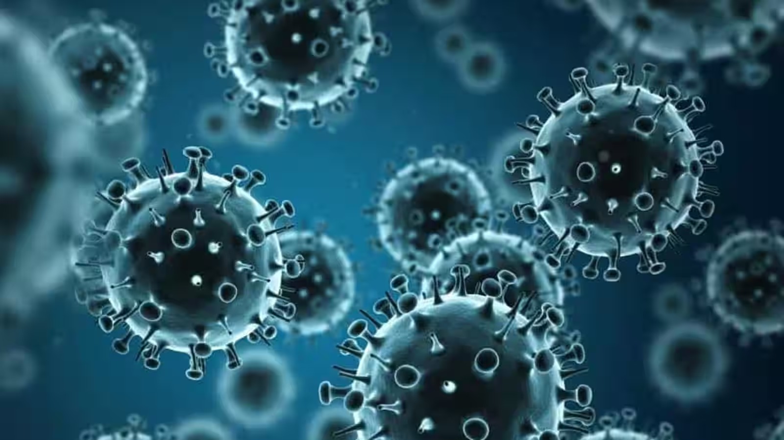
Ahead of Holi, a new virus H3N2 has become a cause of concern in India with its rapid spread. Here are precautions you must follow during the festival of colours to prevent infection.

Holi celebrations have already begun in many parts of the country and the preparations for festival special food menu from gujiya, malpua, kheer to dahi vada as well as shopping for gulal, abeer, pichkari, are on in full swing. While Holi is all about keeping your everyday stress aside and soaking in the festive and colourful spirit of the festival, a new virus H3N2 has become a cause of concern in India with its rapid spread. It is thus important to not only take care of your skin and hair during the festival, but also follow social distancing measures to prevent from the infections. If you suffer from any of the symptoms from fever, respiratory symptoms like cough and runny nose, or body aches, nausea, vomiting, or diarrhoea, you must consult your doctor and start medication.

“Skin and hair issues in the festival of colours are not going to be the only concern this year as a new virus H3N2 has been spreading all over India, due to the rapid shift in weather from cold to warmer temperatures. The increase in the number of patients complaining of viral infections and chest congestion demonstrates how seasonal change is having a negative influence on people’s health. Pollution is also playing a significant influence in increasing the number of patients affected by viral diseases. With Holi coming into the picture, the elderly, children, and pregnant women are the most vulnerable to infection. As a result, they must exercise extreme caution while celebrating Holi. Apart from asthmatic patients, there are many additional individuals who have serious lung infections and are having difficulty breathing they need to be extra careful as they can inhale the colours which can cause serious harm to their health,” says Dr. SK Chhabra, Head of Department – Pulmonary, Primus Hospital, New Delhi.
H3N2 influenza in India; tips to prevent infection while playing Holi

“During Holi, even small respiratory problems must be reported to a pulmonologist or a physician to avoid the condition from worsening. Similar to seasonal flu virus H3N2, other viruses are also in circulation, including H1N1 and adenoviruses and these infections can cause fever, respiratory symptoms like cough and runny nose, as well as other symptoms including body aches, nausea, vomiting, or diarrhoea,” adds Dr. SK Chhabra.

Keeping skin and hair concerns in mind Dr. Navya Handa, Consultant Dermatologist and Cosmetologist at Primus Super Specialty Hospital suggests some useful tips such as a few hours before playing Holi, you should apply oil to your body and hair – you may use coconut or almond, or olive oil. “In addition, you should apply a good sunscreen on your face and any exposed skin. It hydrates the skin and reduces the number of colours absorbed by the skin and hair. Tie your hair instead of keeping them open so that there is less space for chemicals to get to the scalp,” says Dr Handa.

PRECAUTIONS TO FOLLOW DURING HOLI FOR PREVENTING FLU SPREAD

These measures will help prevent the flu and limit its spread:

1. Wash your hands frequently: Always and extensively clean your hands with soap and water, or an alcohol-based hand sanitizer if neither is available.

2. Cover your coughs and sneezes: Use a tissue or your elbow to sneeze or cough into if you must expose others to your germs. Do a quick handwashing.

3. Don’t touch your face unnecessarily: Don’t touch your face too much; that includes your eyes, nose, and mouth.

4. Clean surfaces: Keep frequently touched surfaces clean to avoid picking up the virus from them and spreading it to your body.

5. Avoid crowd: If at all possible, avoid congested areas. Keep your distance from those who are ill. You should avoid swine barns at seasonal fairs and elsewhere if you are at high risk of problems from the flu because of your age (younger than 5 or 65 or older), your health (pregnant or have a chronic illness like asthma).

Read more at-https://bit.ly/3ISDncl
Categories
COVID-19 Government Guidelines Medical Equipments Medical Updates

Paxlovid reduced hospitalisation, death risk during Omicron wave: Study

Antiviral drug, paxlovid, significantly reduced the likelihood of hospitalisation or death from COVID-19 in people at risk of severe illness during the Omicron wave, according to a study.

Antiviral drug, paxlovid, significantly reduced the likelihood of hospitalisation or death from COVID-19 in people at risk of severe illness during the Omicron wave, according to a study. The research, published in the Canadian Medical Association Journal, aimed to evaluate the effectiveness of paxlovid, a combiantion of nirmatrelvir and ritonavir drugs, in preventing severe illness during the emergence of the Omicron variant.

The researchers from Public Health Ontario looked at data on adults with mild disease in Ontario, Canada who tested positive for SARS-CoV-2 by polymerase chain reaction (PCR) test between April 4 and August 31, 2022.

They compared 8,876 patients treated with nirmatrelvir–ritonavir with 1,68, 669 who were not treated. Most patients were older than 70 years, were vaccinated and had potential drug–drug interactions.

A previous randomised controlled trial conducted before the emergence of the Omicron variant had found nirmatrelvir–ritonavir to be effective at treating patients.

However, that trial did not include people who had been vaccinated or who had potential drug–drug interactions.

Paxlovid reduced hospitalisation, death risk during Omicron wave: Study

“Our study, in conjunction with previous clinical trials and observational research, supports the effectiveness of nirmatrelvir–ritonavir at reducing hospital admission from COVID-19 and all-cause death,” said study lead author Kevin Schwartz from Public Health Ontario.

The researchers found that for every 62 people treated with nirmatrelvir–ritonavir, the medication prevented one case of severe COVID-19.

“This study highlights the importance of testing for SARS-CoV-2 if you have symptoms, and access to Paxlovid for those at risk for severe COVID-19,” Schwartz said.

“If you test positive for COVID-19, are over 60 years of age, or if you have other risk factors for severe infection, such as chronic medical conditions or are undervaccinated, contact your health care provider or pharmacy within 5 days of symptoms starting and ask about Paxlovid,” the scientist added.

Read more at-https://bit.ly/3YxzfFe

Categories
Government Guidelines Medical Equipments Medical Updates

Health expert on how Mechanical Thrombectomy is extremely beneficial for stroke patients

Currently, stroke cases are rising at a rapid rate in India and it is commonly seen in elderly people but there are rare instances of youngsters getting affected too. Did you know that mechanical thrombectomy can be helpful to recover from a stroke?

stroke, or brain attack, happens when blood flow to your brain is reduced or interrupted so, health experts recommend B.E.F.A.S.T (Balance Loss, Eyesight Blurring or Loss, Facial drooping, Arm weakness, Speech difficulties and Time) treatment within the golden hour of the stroke to recover without any severe complications. Currently, stroke cases are rising at a rapid rate in the country where stroke is commonly seen in elderly people but there are rare instances of youngsters getting affected too.

Moreover, it can lead to higher mortality and morbidity rates all over the world but did you know that mechanical thrombectomy can be helpful to recover from a stroke? In an interview with HT Lifestyle, Dr Pavan Pai, Consultant Interventional Neurologist at Wockhardt Hospital on Mira Road, explained, “Mechanical Thrombectomy is a type of interventional procedure that is extremely beneficial for stroke patients. This procedure will help to remove a blood clot from the artery that supplies blood to the brain. Speak to your doctor about it. Remember that timely intervention is beneficial for stroke patients.”

Health expert on how Mechanical Thrombectomy is extremely beneficial for stroke patients

Highlighting that a stroke can affect the mobility of the patient, he elaborated, “Many patients get paralysed after a stroke. If an emergency like a brain stroke occurs, know that in a large artery occlusion after IV thrombolysis within window period of 4.5 hours or when a TPA cannot be opted for due to recent surgery or outside window period situation then mechanical thrombectomy is a safe option that is preferred by the Doctors. Put in simple words, mechanical thrombectomy is one of the safest options to manage brain stroke by removing clots that clog the arteries that are responsible for supplying blood to the brain. Thus, this procedure is helpful for patients with acute ischemic stroke i.e. brain stroke victims as the brain does not get blood supply as required. Are you aware? The blood vessels in the brain become narrow because of the fats or plaque.”

He advised, “Diagnostic techniques such as CT or MRI scans can help to know about the blockage of the artery and then only the doctor will be able to decide if the patient is fit to undergo, mechanical thrombectomy. For good clinical outcomes Mechanical thrombectomy has to be done within 6 hours of the onset of a stroke in front arteries and upto 12 hours in arteries supplying back of the brain. In certain situations it can be done upto 24 hours. This process means recanalization or re-opening and restoration of normal blood flow which is done via groin or wrist arteries.”

Dr Pavan Pai revealed, “During this procedure, an aspiration catheter or stent retriever or sometimes both are advanced into the site of the occlusion. There is X-ray imaging is used to check if the stent goes past the clot, clears the clot, and resumes the blood flow. The hospitals should be well-equipped to perform this procedure under the guidance of a skilled expert. The impact of this surgery is immediate and many patients become disability free after the procedure. Those who don’t show immediate recovery show better functionality at the end of 3 months than their counterparts who hadn’t undergone the procedure in the window period initially.”

Read more at-https://bit.ly/40MLIpU

Categories
Medical Updates Medicine & Health

Why we should not reuse plastic water bottles

While there is not yet any clear evidence between microplastics and serious illnesses, researchers are increasingly concerned over their long-term effects on our bodies.

Taking a swig from your plastic water bottle will do more than slake your thirst — you will also be gulping down an unhealthy dose of microplastics — tiny plastic particles measuring less than 5mm. Being plastic, these particles do not decompose all that easily and will accumulate over time in our bodies — a process called bioaccumulation.

While there is not yet any clear evidence between microplastics and serious illnesses, researchers are increasingly concerned over their long-term effects on our bodies.

Key to this concern are the chemicals used in the manufacture of plastics, some of which have already been linked to serious diseases.

The presence of microplastics in human stool suggests we are exposed to microplastics in our daily lives.

They have found their way into the food chain, raising concerns about food safety.

They are also present in bottled water globally.

Why we should not reuse plastic water bottles

Studies investigating microplastics in bottled water have reported particle sizes of less than 1mm in most, released from the bottle material, the bottleneck and the cap.

The colour of the particles coming from the bottle material itself is transparent while those originating from the caps are either blue or green.

The highest detected plastics polymer is polyethylene terephthalate (PET) which is used to manufacture both the bottle material and cap.

Studies also provide evidence that microplastic in bottled water is due to multiple factors such as physical stress during transport, bottle shaking and high-pressure water injection into the bottles at the production plants.

Additionally, thermal impact during storage also worsens the fragmentation process.

Reusable PET water bottles have higher microplastic particles than single-use PET bottles.

Frequent opening and closing of the bottles also cause more particles to be formed due to friction.

The critical question remains unanswered: to what extent do microplastic particles found in bottled water threaten human health?

Researchers have developed a number of hypotheses on both the physical and chemical hazards.

No published study has directly studied the impact of plastic particles on humans.

The only existing research relies on laboratory tests that expose cells or human tissues to microplastics or those that employ rodents.

According to the World Health Organisation, only microplastics with particle sizes smaller than 1.5 μm (1.5 micrometres) can be ingested or absorbed due to their solubility and excreted directly.

Thus ingested microplastic particles (<1.5 μm) from bottled water are capable of migrating through the intestinal wall and reaching various body tissues, including the gut, liver and lymph nodes.

Minute particles (<1.5 μm) which enter cells or tissues might irritate just by being a foreign presence causing lung tissue inflammation which could lead to cancer.

The accumulation of these particles in human tissues has been linked with chemical toxicity.

Compounds such as plasticisers, stabilisers and pigments used in the production can be released by microplastics and travel through our bodies in the bloodstream.

These chemicals have been linked to health problems such as inflammation, genotoxicity, oxidative stress and damage to the gastrointestinal tract.

Chemicals released from bottled water packaging materials are now known as emerging pollutants and endocrine-disrupting chemicals (EDCs) that can cause serious health problems including cancer and developmental defects.

The long-term effects of microplastics exposure on human health are not yet fully understood and research is progressing.

But it is clear they are a potential hazard and steps should be taken to limit our exposure to them in daily life.

In the event you use bottled water as your primary source of drinking water, you should make an effort to minimise the shaking movements of the bottles and also unnecessarily opening and closing the bottle.

And it is not recommended to reuse plastic water bottles.

Reuse increases the rate of inner surface abrasion, releasing additional microplastic particles from the inner surface of the bottle.

It is also essential to store bottles in a cool and dry place to minimise their exposure to heat and sunlight. Sunlight can accelerate bottle degradation — they become more brittle and fragile — which leads to more microplastic particle release.

Additionally, the heat also causes these PET bottles to leach chemical pollutants such as plasticisers which can contaminate the water.

These chemicals, such as phthalates and bisphenol A (BPA) are harmful to human health if consumed in large amounts.

Read more at-https://bit.ly/3I9mifc

Categories
COVID-19 Government Guidelines Medicine & Health

World Cancer Day: High-grade vs low-grade cancer; what’s the difference, symptoms, treatment

World Cancer Day 2023: While low grade tumours spread slowly, high-grade malignancies are aggressive and necessitate immediate treatment to prevent disease from spreading to other parts of the body. All you want to know.

 Cancer is caused by changes to our genes that control the way our cells function. Genetics, environmental factors, lifestyle factors can all affect your chances of having cancer. Some cancers are inherited which means you may be born with a predisposition to cancer. However, most of the cancers occur due to environmental and lifestyle reasons. Our bodies are made up of trillions of cells and the healthy body function is maintained with old cells dying and new cells replacing them. However, in case of cancer, the DNA of a cell gets damaged and there are too many of such abnormal cells which are beyond immune system’s control. These abnormal cells form tumour and in many cases they spread to other body parts if not treated. Some of the cancers are fast growing or high grade, others are slow growing or low grade. Cancer cells that have more genetic damage grow faster than those will less damage.

Stages of cancer

While low grade tumours spread slowly, high-grade malignancies are aggressive and necessitate immediate treatment to prevent disease from spreading to other parts of the body.

“Cancer is a broad category of disorders that all have one thing in common: they all occur when normal cells transform into cancer cells that multiply and spread. For most malignancies, the stage is a Roman numeral ranging from I to IV, with IV being the most advanced and indicating that cancer has spread. Stages are sometimes further classified by letters such as A and B,” says Dr Anil Kumar MD (Internal Medicine) (PGIMER), DM (Medical Oncology and Hematology), Consultant Medical Oncologist, Jindal Naturecure Institute, Bangalore.

World Cancer Day: High-grade vs low-grade cancer; what's the difference, symptoms, treatment

Difference between high-grade and low-grade cancer

Cancer cells that look like healthy cells and tissue are low grade tumours while those that look abnormal in appearance are high grade; they spread more rapidly.

Cancer cells in high-grade (poorly differentiated) tumours seem considerably different from normal cells. High-grade malignancies typically have a good response to therapy due to high cell multiplication rate than low-grade tumours and necessitate immediate treatment to prevent the disease from spreading to other parts of the body. In contrast, cancer cells in low-grade (well-differentiated) tumours resemble cells in normal tissue. In general, these tumours spread slowly. Most cancer grades are based on how aberrant the cancer cells appear under a microscope. This is known as differentiation,” adds Dr Anil.

Symptoms

“The grade is significant because tumours with more abnormal-looking cells develop and spread more quickly. Painless lump, change in bowel habits or blood in stools, constipation, non-healing ulcer, bloody nipple discharge, excessive bleeding per vagina, change in voice, difficulty swallowing, foreign body sensation in the throat, change in the appearance of a mole, fatigue, weakness, loss of appetite, weight changes are the most common symptoms of both types of cancer,” says Dr Anil.

Prevention, diagnosis and treatment

Screening is one way, while early diagnosis is another. It is defined as the presumed detection of undetected disease in a population of persons who appear healthy and asymptomatic utilising tests, examinations, or other methods that may be provided to the target population rapidly and cheaply.

“A screening programme must involve the full screening process, from inviting the target group to providing access to effective care for those who are diagnosed with the disease. Cancer screening tests can help in the early detection of the disease, which raises the likelihood of a successful course of treatment,” adds the expert.

Read more at-https://bit.ly/3Ys7Dkx

Categories
COVID-19 Government Guidelines Medical Equipments Medical Updates

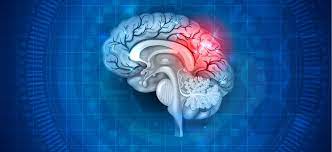
Long Covid might teach us how to prevent Alzheimer’s

Researchers are uncovering links between viruses and brain diseases, which could point to a whole new approach to treatment.

Science is starting to uncover an unnerving fact about viruses: Some might affect our brains over the long haul. It came as a shock that SARS-CoV-2 can lead to lingering neurological problems — a post-viral syndrome we call long Covid. But the phenomenon might not be unique to this virus.

Scientists are finding links between common viruses such as influenza and brain diseases like multiple sclerosis, Parkinson’s, Alzheimer’s and ALS. Researchers are hoping that identifying a viral link might finally allow scientists to figure out what causes these mysterious, deadly ailments and develop new treatments.

A new study, published last week and summarized in Science, used a vast trove of electronic medical records to find thousands of people with neurodegenerative diseases and tease out correlations with 22 different kinds of infections. The biggest viral risk factor for dementia was encephalitis, which is an infection of the brain often caused by a mosquito or tick-borne disease. Other viruses tied to dementia included influenza, herpes zoster (shingles) and HPV.

Earlier studies had looked for specific viral links to Alzheimer’s and found correlations with herpes and some forms of HPV. And a study published last year showed that Epstein-Barr virus was necessary for developing MS. But scientists still haven’t figured out exactly what role the viruses play — whether they’re a direct trigger or have some peripheral role. Almost everyone on the planet carries Epstein-Barr virus, which causes mononucleosis, but only a tiny fraction ends up getting MS.

To better understand how viruses might be affecting the brain, I stopped by the National Institutes of Health to visit Avindra Nath, who is one of the few neurologists specializing in viruses — an interest he picked up after treating AIDS patients in the early 1980s. In 2014, he was the first neurologist to travel to Liberia to treat Ebola patients, and he said a fraction of those who recover suffer neurological symptoms similar to long Covid — especially chronic fatigue.

He told me that what really interests him are viruses that are embedded in our genetic codes — called endogenous retroviruses. They may have snuck in through a sexually transmitted virus that works itself into an embryo. These viruses are a source of genetic variation — and can add novel DNA to our genomes. Like mutations, these occasionally incur an advantage and spread through the population. About 8% of our genome is made of these embedded viruses.

Long Covid might teach us how to prevent Alzheimer’s

Nath’s interest in these started when he was treating a patient who had both HIV and ALS, and after taking antiretroviral drugs, the ALS disappeared. A series of human and animal studies convinced Nath that ALS might sometimes be triggered by an embedded virus called HERV-K.

Normally HERV-K is active during fetal development, so it may have spread through the human population because it does something useful in utero. But after we’re born, it normally shuts off (due to external chemical switches that bind to DNA).

Sometimes, he told me, these “off switches” fail, and one of these embedded viruses can become reactivated. That failure might be part of the cause of ALS. After years of animal studies, he started preliminary human trials assessing the effects of antiviral drugs on patients with ALS. He’s now planning a placebo-controlled clinical trial for drugs that he’s shown can suppress HERV-K.

He’s also studying the kinds of viruses we catch from the outside world. He thinks viruses are more likely to trigger brain diseases indirectly — not by getting directly into neurons, but by prompting inflammation. It’s the immune system’s response that causes the problem.

He’s now studying people with neurological problems following Covid infection — something that he estimates affected about 10% of people infected before vaccines were available and a much smaller fraction today. He said that at first he was very skeptical of claims that the virus hides out in the brain, but more recent studies have made him take it seriously, especially autopsy studies done by Dan Chertow at the NIH. It might be that those residual traces of the virus are causing persistent inflammation.

It’s possible that this has always been happening to some fraction of people who pick up common viruses. It’s just that it didn’t get all that much attention before the Covid pandemic. Nath said virology and neurology were two completely different worlds, and he was unique in trying to navigate both fields at the same time.

If viruses do indeed play a role in some of our most feared and costly diseases, that could point to a whole new approach to treatment and prevention. In the future, everyone might get vaccinated against Epstein-Barr virus as a protection against MS. And it may turn out that you can reduce your odds of getting Alzheimer’s disease by getting that shingles shot and a flu shot every year. If HPV adds to the risk, it could motivate the medical community to redouble its efforts to get that vaccine to more people.

Scientists could even eventually develop a universal, variant-proof Covid vaccine that would finally prevent the risk of long Covid.

Someday, people may look back at all the expensive drugs we tested for dementia and other brain diseases and wonder how we overlooked the benefits of cheap vaccines for so long.

Read more at-https://bit.ly/3kGNokj

Categories
COVID-19 Government Guidelines Medical Updates Medicine & Health

Practising gratitude can help you reduce stress: Research

Gratitude has a special stress-buffering effect on responses to and recovery from acute psychological stress, which can help to enhance cardiovascular health, according to a study done by researchers from Irish universities with 68 adults.

Researchers from Irish institutions discovered that gratitude has a unique stress-buffering effect on both reactions to and recovery from acute psychological stress, which can contribute to better cardiovascular health. Knowing that stress affects humans and has an impact on their health and well being, specifically causing high blood pressure and increasing cardiovascular morbidity and coronary heart disease, it is critical to understand our reactions to stress and determine whether there are any factors that can play key stress-buffering roles.

In the article “Gratitude, affect balance, and stress buffering: A growth curve examination of cardiovascular responses to a laboratory stress task”, published in January in the Journal of Psychophysiology, Brian Leavy, Brenda H. O’Connell and Deirdre O’Shea propose that, although previous research suggest that gratitude and affect-balance play key stress-buffering roles, to date little has been known about the impact of these variables on cardiovascular recovery from acute psychological stress.

That was the focus of the study by the researchers from the Universities of Maynooth and Limerick in Ireland, who also sought to find out whether affect balance moderates the relationship between gratitude and cardiovascular reactions to acute psychological stress.

The research carried out at the Irish University of Maynooth involved 68 undergraduate students (24 male and 44 female), aged between 18 and 57 years. This study used a within-subjects experimental design with lab tasks in which stress was induced to participants and then cardiovascular reactivity and recovery in response to this was measured.

Practising gratitude can help you reduce stress: Research

The results showed that state gratitude predicted lower systolic blood pressure responses throughout the stress-testing period, which means that the state of gratitude has a unique stress-buffering effect on both reactions to and recovery from acute psychological stress. It was also found that affect balance amplifies the effects of state gratitude.

These findings have clinical utility as there are several low-cost gratitude interventions which can contribute to well-being (Wood et al., 2010). For example, previous research has shown how cardiac patients who make use of gratitude journals have better cardiovascular outcomes than those who do not (Redwine et al., 2016).

Combined with the results of this study and previous work, gratitude may thus constitute a useful point of intervention for the improvement of our cardiovascular health.

Read more at-https://bit.ly/3Rb4u6f

Categories
COVID-19 Government Guidelines Medical Equipments Medical Updates Medicine & Health

High fat diet could reduce ability to regulate food intake: Study

Understanding the brain’s role and the complex mechanisms that lead to overeating, a behaviour that can lead to weight gain and obesity, could help develop therapies to treat it.

Regularly eating a high-fat/calorie diet could reduce the brain’s ability to regulate calorie intake. New research in rats found that after short periods of being fed a high-fat/high-calorie diet, the brain adapts to react to what is being ingested and reduces the amount of food eaten to balance calorie intake.

The researchers from Penn State College of Medicine, US, suggest that calorie intake is regulated in the short-term by cells called astrocytes (large star-shaped cells in the brain that regulate many different functions of neurons in the brain) that control the signalling pathway between the brain and the gut. Continuously eating a high-fat/calorie diet seems to disrupt this signalling pathway. The study was published in The Journal of Physiology.

Understanding the brain’s role and the complex mechanisms that lead to overeating, a behaviour that can lead to weight gain and obesity, could help develop therapies to treat it. Obesity is a global public health concern associated with an increased risk of cardiovascular diseases and type 2 diabetes. In England, 63 per cent of adults are considered above a healthy weight and around half of these are living with obesity. One in three children leaving primary school is overweight or obese1.

Dr Kirsteen Browning, Penn State College of Medicine, US, said,

“Calorie intake seems to be regulated in the short-term by astrocytes. We found that a brief exposure (three to five days) of a high fat/calorie diet has the greatest effect on astrocytes, triggering the normal signalling pathway to control the stomach. Over time, astrocytes seem to desensitise to high-fat food. Around 10-14 days of eating a high fat/calorie diet, astrocytes seem to fail to react and the brain’s ability to regulate calorie intake seems to be lost. This disrupts the signalling to the stomach and delays how it empties.”

High fat diet could reduce ability to regulate food intake: Study

Astrocytes initially react when high-fat/calorie food is ingested. Their activation triggers the release of gliotransmitters, chemicals (including glutamate and ATP) that excite nerve cells and enable normal signalling pathways to stimulate neurons that control how the stomach works. This ensures the stomach contracts correctly to fill and empty in response to food passing through the digestive system. When astrocytes are inhibited, the cascade is disrupted. The decrease in signalling chemicals leads to a delay in digestion because the stomach doesn’t fill and empty appropriately.

The vigorous investigation used behavioural observation to monitor food intake in rats (N=205, 133 males, 72 females) which were fed a control or high fat/calorie diet for one, three, five or 14 days. This was combined with pharmacological and specialist genetic approaches (both in vivo and in vitro) to target distinct neural circuits. Enabling the researchers to specifically inhibit astrocytes in a particular region of the brainstem (the posterior part of the brain that connects the brain to the spinal cord), so they could assess how individual neurons behaved to studying rats’ behaviour when awake. (ANI)

Human studies will need to be carried out to confirm if the same mechanism occurs in humans. If this is the case, further testing will be required to assess if the mechanism could be safely targeted without disrupting other neural pathways.

The researchers have plans to further explore the mechanism. Dr Kirsteen Browning said,

“We have yet to find out whether the loss of astrocyte activity and the signalling mechanism is the cause of overeating or that it occurs in response to the overeating. We are eager to find out whether it is possible to reactivate the brain’s apparent lost ability to regulate calorie intake. If this is the case, it could lead to interventions to help restore calorie regulation in humans.”

Read more at-https://bit.ly/3HChfmU

Categories
COVID-19 Government Guidelines Medical Equipments Medical Updates Medicine & Health Techniques

Covid infection could damage foetuses of pregnant women: Study

Foetuses and placentas of pregnant women, especially those infected with COVID-19 at earlier points in the pandemic, were found to bear a greater risk of experiencing growth impairment or vascular lesions in organs and brain, according to a new research.

Foetuses and placentas of pregnant women, especially those infected with COVID-19 at earlier points in the pandemic, were found to bear a greater risk of experiencing growth impairment or vascular lesions in organs and brain, according to a new research.

According to the research, results demonstrate that the different strains of the virus that emerged during the pandemic led to varying degrees of damage, which was especially higher by those involving pre-Omicron variants. The detected placental lesions could potentially harm both development and health in some of the affected unborn children, the study said.

Using prenatal magnetic resonance imaging, a group of Medical University of Vienna, Austria, researchers examined the placentas and foetuses of women who tested positive for SARS-CoV-2 during pregnancy, the study said.

The research was published in the journal The Lancet Regional Health – Europe. While such damage occurs more infrequently and is less severe with the currently circulating Omicron sub-lineages, the study authors still advocate early detection measures for pregnant women who test positive for coronavirus.

Contrary to previous studies, in which SARS-CoV-2-related abnormalities were only identified postnatally and/or through histopathological procedures, the research team focused on prenatal imaging findings, the study said.

Using prenatal magnetic resonance imaging (MRI), 76 scans of placentas and foetuses of pregnant women were performed in the study: 38 following a confirmed SARS-CoV-2 infection (pre-Omicron or Omicron variants) and 38 in healthy control cases, the study said.

The study found that the placentas in both the pre-Omicron and the Omicron groups revealed abnormalities.

Covid infection could damage foetuses of pregnant women: Study

“Infections with pre-Omicron variants, such as Delta, led to significantly greater damage in the form of vascular events such as blood clots or bleeding than with the Omicron subvariants currently circulating through the population,” noted lead author Patric Kienast, outlining a key finding of the study.

The researchers attribute the varying extent of damage to the placenta caused by different virus strains to the fact that Omicron sub-lineages are less likely than their predecessors to result in severe cases, and to higher vaccination rates as the pandemic has progressed, the study said.

“In any case, our results showed that the two non-vaccinated study participants developed placental abnormalities following infection with SARS-CoV-2 Omicron, compared with just one out of six of the women who had received three doses of the vaccine,” confirmed senior author Gregor Kasprian.

Oxygen, nutrients and metabolic products are exchanged between mother and child in the placenta. Attached to the uterine wall, this organ forms such a strong barrier against the coronavirus that only 3 per cent or fewer of foetuses whose mothers test positive for SARS-CoV-2 are also infected, the study said.

But as the study shows, the placenta itself is not spared from complications caused by Covid-19. Subsequently, some unborn babies experience stunted growth or bleeding in the brain, the study said.

“This is why the placentas of pregnant women who have been infected with SARS-CoV-2 should be examined as soon as possible after testing positive using prenatal imaging techniques,” advised Daniela Prayer from Medical University of Vienna, citing the importance of scanning, particularly in the case of possible future coronavirus variants with mechanisms similar to Delta, for example.

By doing so, there is a chance to take measures to safeguard the health of the foetus in a worst case scenario, the study said.

Read more at-https://bit.ly/3XAOtZF

Categories
COVID-19 Government Guidelines Medical Equipments Medical Updates Medicine & Health

Male and female hearts respond differently to stress hormone: Study

Male and female hearts react differently to the stress hormone noradrenaline, according to a recent study published in Science Advances.

Male and female hearts react differently to the stress hormone noradrenaline, according to a recent study published in Science Advances. The research on mice may have ramifications for human heart conditions like arrhythmias and heart failure as well as how various sexes react to certain drugs.

The team built a new type of fluorescence imaging system that allows them to use light to see how a mouse heart responds to hormones and neurotransmitters in real-time. The mice were exposed to noradrenaline, also known as norepinephrine. Noradrenaline is both a neurotransmitter and hormone associated with the body’s “fight or flight” response.

The results reveal that male and female mouse hearts respond uniformly at first after exposure to noradrenaline. However, some areas of the female heart return to normal more quickly than the male heart, which produces differences in the heart’s electrical activity.

Male and female hearts respond differently to stress hormone: Study

“The differences in electrical activity that we observed are called repolarization in the female hearts. Repolarization refers to how the heart resets between each heartbeat and is closely linked to some types of arrhythmias,” said Jessica L. Caldwell, first author of the study. Caldwell is a postdoctoral scholar in the UC Davis School of Medicine Department of Pharmacology.

“We know that there are sex differences in the risk for certain types of arrhythmias. The study reveals a new factor that may contribute to different arrhythmia susceptibility between men and women,” Caldwell said.

Heart disease is the leading cause of death for both men and women in the United States. It accounted for about 1 in every 4 male deaths and 1 in every 5 female deaths in 2020. Despite the impact on both sexes, cardiology research has largely been performed on male subjects.

In this study, the researchers were interested in looking at factors that may contribute to arrhythmias. Arrhythmias are a type of heart disorder where the electrical impulses that control heartbeats don’t function properly. They affect somewhere between 1.5% to 5% of the population.

The researchers had not planned to study sex-based responses, according to Crystal M. Ripplinger, senior author of the study. But the researchers started seeing a pattern of different reactions, which led them to realize the differences were sex-based.

Ripplinger, an electrical and biomedical engineer, is a professor in the Department of Pharmacology. When she started her lab at the UC Davis School of Medicine over a decade ago, she exclusively used male animals. That was the norm for most research at the time. But several years ago, she began including male and female animals in her studies.

“Sometimes the data between the two sexes is the same. But if the data start to show variation, the first thing we do is look at sex differences. Using both male and female mice has revealed clues into differences we would never have suspected. Researchers are realizing you can’t extrapolate to both sexes from only studying one,” Ripplinger said.

She notes that with the current study, it’s not clear what the differences in cAMP and electrical activity may mean. “The response in the female mice may be protective — or it may not. But simply documenting that there is a measurable difference in the response to a stress hormone is significant. We are hoping to learn more in future studies,” Ripplinger said.

Read more at-https://bit.ly/3XVhEXd